Loading content - please wait...
Funnelweed pass unnoticed
Funnelweed, though found worldwide, lives in limited locations in Gorontalo. Divers certainly pass by without noticing. But scientists are researching it.
Delicate scalloped algae
Although funnelweed is a member of the brown algae family, its color here in Gorontalo is not brown. It grows in some sandy patches or substrate among other algae. Its color is pale cream. Sometimes, it has tinges of light green.

It grows in beautiful, radiating ribbons. Opaque bands alternate with more transparent ones. Often, this algae forms scallops that curve inwards. On the outer edges and surface sprout almost transparent filaments.
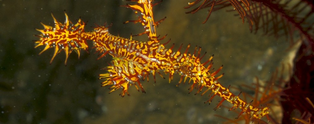
Divers, who take the time to notice funnelweed, quickly appreciate its unique beauty. However, they should not brush against it with unprotected skin. Something about these algae here leaves itchy stings.
Funnelweed around the World
In Gorontalo, this beautiful plant only grows in a few small areas. A few sandy patches are full of these algae. In a couple of other sites, it grows among other algae.
However, it is found worldwide. This includes the Caribbean and Atlantic oceans, as well as the Indian and Pacific ones. The detailed list of locations is quite long. The scientific name for funnelweed is Padina gymnospora. Since it lives in so many oceans, scientist the world over are researching its potential value.
Scientific Research on Padina gymnospora
One group has extracted sulfated polysaccharides from it. Their research indicates anti-inflammatory properties. They used mice in their studies prior to potential use on humans.

Other researchers discovered anti-amyloidogenic agents in funnelweed. This extract in an important component in drugs that combat Alzheimer’s disease.
Field research on Padina gymnospora has found significant absorbent properties in the algae. A synthesized compound from it is over 80% effective in absorbing industrial dyes. Removing these dyes from industrial waste is a priority to build a cleaner world. Using this extract is clean and green solution in reusing industrial wastewater.
Padina gymnospora produces an aragonite calcium carbonate compound as well as phlorotannins. These are found on the algae surfaces. These compounds play an important role in defending the algae from worms and snails.
That chemical protection has no effect on Green turtles, which are herbivores. Padina gymnospora is actually edible. Naturally, it contains no heavy metals or toxins. Although no one really uses funnelweed for human food, it has potential for as a natural fertilizer.
In Gorontalo, only staff of Miguel’s Diving will know where to find these beautiful algae. To see for yourself, please make your dive reservations directly with us.